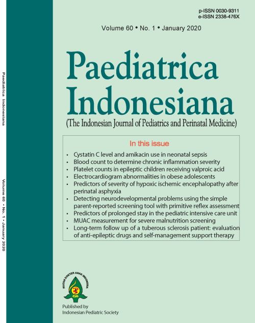
Umbilical arterial profiles as predictors of severity of hypoxic ischemic encephalopathy after perinatal asphyxia
DOI:
https://doi.org/10.14238/pi60.1.2020.24-30Keywords:
perinatal asphyxia; pH; lactate; bace deficit; HIE; umbilical cordAbstract
Background: Perinatal hypoxic-ischemic encephalopathy (HIE) remains a major cause of neurodevelopmental impairment. Umbilical cord blood analysis provides an objective assessment of newborn metabolic status. Accordingly, it is recommended that physicians attempt to obtain venous and arterial samples when there is high risk of neonatal compromise.
Objective To compare the predictive value of umbilical arterial blood pH, lactate and base deficit for subsequent development of severity of hypoxic ischemic encephalopathy (HIE) after perinatal asphyxia and comparison of these parameters to determine which one is superior in predicting severity.
Methods Umbilical cord arterial blood of newborns with perinatal asphyxia was tested for pH, lactate, and base deficit estimation. These newborns were evaluated in level III NICU and divided into two groups. Group 1 had no or signs and symptoms of HIE I and group 2 had signs and symptoms of HIE II/III. Values of pH, lactate, and base deficit were tabulated and analyzed by receiver-operating characteristic curves. Optimal cut-off values were estimated based on the maximal Youden index.
Results Mean pH was significantly lower in group 2 than in group 1, while lactate and base deficit were significantly higher in group 2 than in group 1. Cut-off points for determining severity of HIE were pH <7.13, lactate >6.89 mg/dL, and base deficit >7 mEq/L. Sensitivity and specificity for these cut-off points were 100% and 91.49% for pH, 100% and 85.11% for lactate, and 82.4% and 91.76% for base deficit, respectively. Predictive abilities of all three parameters were similar in determination of HIE severity.
Conclusion Umbilical arterial pH, lactate, and base deficit have excellent accuracy to predict the severity of HIE. All three parameters have similarly good predictive ability.
References
2. Bhimte B, Vamne A. Metabolic derangement in birth asphyxia due to cellular injury with reference to mineral metabolism in different stages of hypoxic-ischemic encephalopathy in Central India. Indian J Med Biochem. 2017;21:86-90. DOI: 10.5005/jp-journals-10054-0027.
3. Wu YW, Backstrand KH, Zhao S, Fullerton HJ, Johnston SC. Declining diagnosis of birth asphyxia in California: 1991–2000. Pediatrics. 2004;114:1584-90. DOI: 10.1542/peds.2004-0708.
4. Black RE, Cousens S, Johnson HL, Lawn JE, Rudan I, Bassani DG, et al. Global, regional, and national causes of child mortality in 2008: a systematic analysis. Lancet. 2010;375:1969-87. DOI: 10.1016/S0140-6736(10)60549-1.
5. ACOG Committee on Obstetric Practice. ACOG Committee Opinion No. 348, November 2006: Umbilical cord blood gas and acid-base analysis. Obstet Gynecol 2006;108:1319–22. DOI: 10.1097/00006250-200611000-00058.
6. Liu L, Johnson HL, Cousens S, Perin J, Scott S, Lawn JE, et al. Global, regional, and national causes of child mortality: an updated systematic analysis for 2010 with time trends since 2000. Lancet. 2012;379:2151-61. DOI: 10.1016/S0140-6736(12)60560-1.
7. Low JA, Panagiotopoulos C, Derrick EJ. Newborn complications after intrapartum asphyxia with metabolic acidosis in the term fetus. Am J Obstet Gynecol. 1994;170:1081–7. DOI: 10.1016/s0002-9378(94)70101-6.
8. King TA, Jackson GL, Josey AS, Vedro DA, Hawkins H, Burton KM, et al. The effect of profound umbilical artery acidemia in term neonates admitted to newborn nursery. J Pediatr. 1998;132:624-9. DOI: 10.1016/s0022-3476(98)70350-6.
9. Sarnat HB, Sarnat MS. Neonatal encephalopathy following fetal distress. A clinical and electroencephalographic study. Arch Neurol. 1976;33:696–705. DOI: 10.1001/archneur.1976.00500100030012.
10. Hanley JA, McNeil BJ. A method of comparing area under receiver operating characteristic curves derived from same cases. Radiology. Radiology. 1983. 148:839-43. DOI: 10.1148/radiology.148.3.6878708.
11. Volpe JJ. Neurology of newborn. Hypoxic-ischemic encephalopathy: biochemical and physiological aspects. 5th ed. 1600 John F. Philadelphia: Saunders Elsevier; 2008. p. 247-50.
12. White CR, Doherty DA, Newnham JP, Pennell CE. The impact of introducing universal umbilical cord blood gas analysis and lactate measurement at delivery. Aust N Z J Obstet Gynaecol. 2014;54:71-8. DOI: 10.1111/ajo.12132.
13. Knutzen L, Svirko E, Impey L. The significance of base deficit in acidemic term neonates. Am J Obstet Gynecol. 2015;213:373.e1-7. DOI: 10.1016/j.ajog.2015.03.051.
14. Georgieva A, Moulden M, Redman CW. Umbilical cord gases in relation to the neonatal condition: the EveREst plot. Eur J Obstet Gynecol Reprod Biol. 2013;168:155-60. DOI: 10.1016/j.ejogrb.2013.01.003.
15. Victory R, Penava D, Da Silva O, Natale R, Richardson B. Umbilical cord pH and base excess values in relation to adverse outcome events for infants delivering at term. Am J Obstet Gynecol. 2004;191:2021-8. DOI: 10.1016/j.ajog.2004.04.026.
16. Shah S, Tracy M, Smyth J. Postnatal lactate as an early predictor of short-term outcome after intrapartum asphyxia. J Perinatol. 2004;24:16–20. DOI: 10.1038/sj.jp.7211023.
17. Huang CC, Wang ST, Chang YC, Lin KP, Wu PL. Measurement of urinary lactate: creatinine ratio for early identification of newborn infants at risk for hypoxic-ischaemic encephalopathy. N Engl J Med. 1999;341:328–35. DOI: 10.1056/NEJM199907293410504.
18. Tuuli MG, Stout MJ, Shanks A, Odibo A, Macones G, Cahill AG. Umbilical cord arterial lactate compared with pH for predicting neonatal morbidity at term. Obstet Gynecol. 2014;124:756-61. DOI 10.1097/AOG.0000000000000466.
19. Wiberg N, Källén K, Herbst A, Olofsson P. Relation between umbilical cord blood pH, base deficit, lactate, 5-minute Apgar score and development of hypoxic ischemic encephalopathy. Acta Obstet Gynecol Scand. 2010;89:1263-9. DOI: 10.3109/00016349.2010.513426.
20. Gjerris AC, Staer-Jensen J, Stener Jorgensen J, Bergholt T, Nickelsen C. Umbilical cord blood lactate: a valuable tool in the assessment of fetal metabolic acidosis. Eur J Obstet Gynecol Reprod Biol. 2008;139:16-20. DOI: 0.1016/j.ejogrb.2007.10.004.
Downloads
Published
How to Cite
Issue
Section
License
Authors who publish with this journal agree to the following terms:
Authors retain copyright and grant the journal right of first publication with the work simultaneously licensed under a Creative Commons Attribution License that allows others to share the work with an acknowledgement of the work's authorship and initial publication in this journal.
Authors are able to enter into separate, additional contractual arrangements for the non-exclusive distribution of the journal's published version of the work (e.g., post it to an institutional repository or publish it in a book), with an acknowledgement of its initial publication in this journal.
Accepted 2020-01-28
Published 2020-01-28


