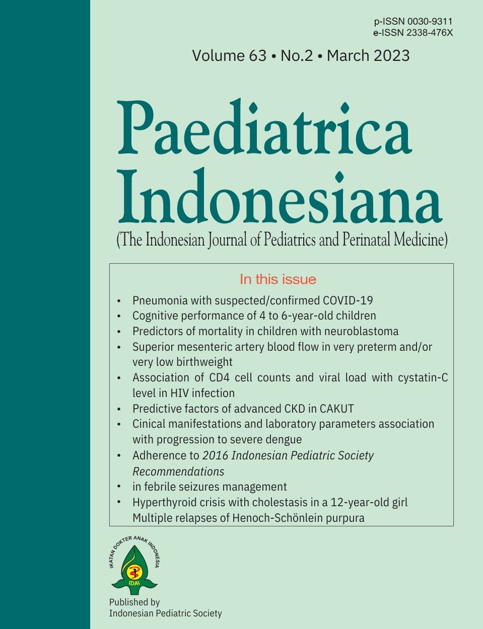
Superior mesenteric artery blood flow in infants of very preterm and very low birthweight and its related factors
Abstract
- Abstract
Background Significant hemodynamic changes in preterm infants during early life could have consequences, especially on the intestinal blood flow. Alteration of superior mesenteric artery (SMA) blood flow may lead to impairment in gut function and feeding intolerance.
Objectives To assess SMA blood flow velocity in very preterm and/or very low birth weight (VLBW) infants in early life and to elucidate the factors influencing them.
Methods This is a cross-sectional study conducted in NICU at Cipto Mangunkusumo Hospital, Jakarta. Superior mesenteric artery (SMA) blood flow was evaluated by peak systolic velocity (PSV), end diastolic velocity (EDV), and resistive index (RI) measurement using Color Doppler US at < 48 hours after birth. Maternal and neonatal data that could be potentially associated with SMA blood flow were obtained. Bivariate analyses were conducted with a P value of < 0.05 considered significant.
Results We examined 156 infants eligible for the study. PSV, EDV, and RI of SMA blood flow were not related to both gestational age and birth weight. Infant with small for gestational age (SGA) showed significantly lower EDV median [15.5 (range 0.0-32.8) vs 19.4 (range 0.0-113.0)] and higher RI [0.80 (range 0.58-1.00) vs 0.78 (range 0.50-1.00)] compared to appropriate for gestational age (AGA). Infants born from mother with preeclampsia showed lower PSV median [(78.2 (range 32.0-163.0) vs 89.7 (range 29.2-357.0)]) and EDV [16.2 (range 0.0-48.5) vs 19.4 (range 0.0-113.0)] compared to without PE, while absent/reverse end-diastolic velocity (AREDV) revealed a lower EDV median [16.9 (range 0.0 – 32.4) vs 19.4 (range 0.0 – 113.0)] compared to no AREDV. Furthermore, infants with hs-PDA showed lower EDV median [16.2 (range 0.0-113.0) vs 19.4 (range 0.0-71.1)] but higher RI median [0.80 (range 0.50-1.00) vs 0.78 (range 0.55-1.00)] compared to non hs-PDA. No difference in SMA blood flow across other factors was observed.
References
2. Maruyama K, Koizumi T, Tomomasa T, Morikawa A. Intestinal blood-flow velocity in uncomplicated preterm infants during the early neonatal period. Pediatr Radiol. 1999;29:472-7 DOI:10.1007/s002470050621.
3. Yin Chia C, Cícero Falcão M, Bueno Cabral JE, de Carvalho Hartmann LG, de Melo Galvão Filho M. Evolution of mesenteric artery blood flow in healthy premature neonates. Nutr Hosp. 2010;25:319 DOI:10.3305/nh.2010.25.2.4641.
4. Kempley ST, Gamsu HR, Vyas S, Nicolaides K. Effects of intrauterine growth retardation on postnatal visceral and cerebral blood flow velocity. Arch Dis Child. 1991;66:1115-8 DOI:10.1136/adc.66.10_spec_no.1115.
5. Martinussen M, Brubakk AM, Vik T, Yao AC. Relationship between intrauterine growth retardation and early postnatal superior mesenteric artery blood flow velocity. Biol Neonate. 1997;71:22-30 DOI:10.1159/000244393.
6. Jain N, Ramji S, Jain A, Modi M, Sharma P. Early prediction of feed intolerance in very low birth weight preterm infants using superior mesenteric artery blood flow velocity. J Matern Fetal Neonatal Med. 2019:1-6 DOI:10.1080/14767058.2019.1591362.
7. Maruyama K, Koizumi T. Superior mesenteric artery blood flow velocity in small for gestational age infants of very low birth weight during the early neonatal period. 2001;29:64-70 DOI:doi:10.1515/JPM.2001.009.
8. Van Bel F, Van Zoeren D, Schipper J, Guit GL, Baan J. Effect of indomethacin on superior mesenteric artery blood flow velocity in preterm infants. J Pediatr. 1990;116:965-70 DOI:10.1016/s0022-3476(05)80662-6.
9. Shimada S, Kasai T, Konishi M, Fujiwara T. Effects of patent ductus arteriosus on left ventricular output and organ blood flows in preterm infants with respiratory distress syndrome treated with surfactant. J Pediatr. 1994;125:270-7 DOI:10.1016/s0022-3476(94)70210-1.
10. Wong SN, Lo RN, Hui PW. Abnormal renal and splanchnic arterial Doppler pattern in premature babies with symptomatic patent ductus arteriosus. J Ultrasound Med. 1990;9:125-30 DOI:10.7863/jum.1990.9.3.125.
11. Havranek T, Rahimi M, Hall H, Armbrecht E. Feeding preterm neonates with patent ductus arteriosus (PDA): intestinal blood flow characteristics and clinical outcomes. J Matern Fetal Neonatal Med. 2015;28:526-30 DOI:10.3109/14767058.2014.923395.
12. Pelicia SMC, Fekete SMW, Corrente JE, Rugolo L. The effect of early-onset preeclampsia on the intestinal blood flow of preterm infants. J Matern Fetal Neonatal Med. 2021;34:2235-9 DOI:10.1080/14767058.2019.1661378.
13. Cetinkaya M, Ozkan H, Koksal N. Maternal preeclampsia is associated with increased risk of necrotizing enterocolitis in preterm infants. Early Hum Dev. 2012;88:893-8 DOI:10.1016/j.earlhumdev.2012.07.004.
14. Havranek T, Ashmeade TL, Afanador M, Carver JD. Effects of maternal magnesium sulfate administration on intestinal blood flow velocity in preterm neonates. Neonatology. 2011;100:44-9 DOI:10.1159/000319049.
15. Wang KG, Chen CY, Chen YY. The effects of absent or reversed end-diastolic umbilical artery Doppler flow velocity. Taiwan J Obstet Gynecol. 2009;48:225-31 DOI:10.1016/s1028-4559(09)60294-1.
16. Yoon BH, Lee CM, Kim SW. An abnormal umbilical artery waveform: a strong and independent predictor of adverse perinatal outcome in patients with preeclampsia. Am J Obstet Gynecol. 1994;171:713-21 DOI:10.1016/0002-9378(94)90087-6.
17. Gonzalez JM, Stamilio DM, Ural S, Macones GA, Odibo AO. Relationship between abnormal fetal testing and adverse perinatal outcomes in intrauterine growth restriction. Am J Obstet Gynecol. 2007;196:e48-51 DOI:10.1016/j.ajog.2007.01.010.
18. Karsdorp VH, van Vugt JM, van Geijn HP, Kostense PJ, Arduini D, Montenegro N, et al. Clinical significance of absent or reversed end diastolic velocity waveforms in umbilical artery. Lancet. 1994;344:1664-8 DOI:10.1016/s0140-6736(94)90457-x.
19. Torrance HL, Mulder EJ, Brouwers HA, van Bel F, Visser GH. Respiratory outcome in preterm small for gestational age fetuses with or without abnormal umbilical artery Doppler and/or maternal hypertension. J Matern Fetal Neonatal Med. 2007;20:613-21 DOI:10.1080/14767050701463662.
20. Hartung J, Kalache KD, Heyna C, Heling KS, Kuhlig M, Wauer R, et al. Outcome of 60 neonates who had ARED flow prenatally compared with a matched control group of appropriate-for-gestational age preterm neonates. Ultrasound Obstet Gynecol. 2005;25:566-72 DOI:10.1002/uog.1906.
Copyright (c) 2023 Evita Karianni Bermanshah Ifran, Wresti Indriatmi, Tetty Yuniarti, Nadjib Advani, Saleha Sungkar, Dewi Irawati Soeria Santoso, Rinawati Rohsiswatmo, Yvan Vandenplas, Badriul Hegar Syarif

This work is licensed under a Creative Commons Attribution-NonCommercial-ShareAlike 4.0 International License.
Authors who publish with this journal agree to the following terms:
Authors retain copyright and grant the journal right of first publication with the work simultaneously licensed under a Creative Commons Attribution License that allows others to share the work with an acknowledgement of the work's authorship and initial publication in this journal.
Authors are able to enter into separate, additional contractual arrangements for the non-exclusive distribution of the journal's published version of the work (e.g., post it to an institutional repository or publish it in a book), with an acknowledgement of its initial publication in this journal.
Accepted 2022-12-12
Published 2023-04-06


