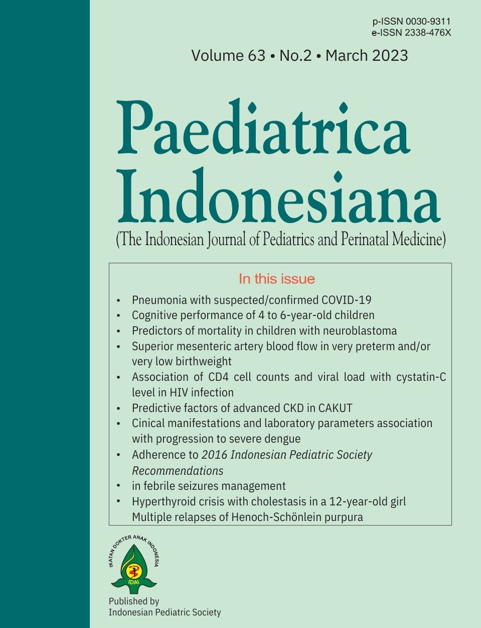
Evaluating the importance of clinical manifestations and laboratory parameters associated with progression to severe dengue in children
DOI:
https://doi.org/10.14238/pi63.2.2023.102-18Keywords:
children, dengue, risk prediction, warning signAbstract
Background The ability to predict the progression to severe dengue is crucial in managing patients with dengue fever. Severe dengue is defined by one or more of the following signs: severe plasma leakage, severe bleeding, or severe organ involvement as it can be a life-threatening condition if left untreated.
Objective To identify clinical manifestations and laboratory parameters associated with dengue hemorrhagic fever disease progression in children by systematic review and meta-analysis.
Methods We searched six medical databases for studies published from Jan 1, 2000, to Dec 31, 2020. The meta-analysis used random-effects or fixed-effects models to estimate pooled effect sizes. We assessed heterogeneity using Cochrane Q and I2 statistics, publication bias by Egger’s test and LFK index (Doi plot), and categorized subgroup analysis by country. This study was registered with PROSPERO, CRD42021224439.
Results We included 49 papers in the systematic review, and we encased the final selected 39 papers comprising 23 potential predictors in the meta-analyses. The other 10 papers were not included because the raw data could not be calculated for the effect measure in the meta-analysis. Among 23 factors studied, seven clinical manifestations demonstrated association with disease progression in children, including neurological signs, gastrointestinal bleeding, clinical fluid accumulation, hepatomegaly, vomiting, abdominal pain, and petechiae. Six laboratory parameters were associated during the early days of illness, including elevated hematocrit, aspartate aminotransferase [AST], and alanine aminotransferase [ALT], low platelet count, low albumin levels, and elevated activated partial thromboplastin time. Dengue virus serotype 2 (DENV-2) and secondary infections were also associated with severe disease progression.
Conclusion This review supports the use of the warning signs described in the 2009 WHO guidelines. In addition, monitoring serum albumin, AST/ALT levels, identifying infecting dengue serotypes, and immunological status can improve the prediction of further risk of disease progression.
References
2. Stanaway JD, Shepard DS, Undurraga EA, Halasa YA, Coffeng LE, Brady OJ, et al. The global burden of dengue: an analysis from the Global Burden of Disease Study 2013. Lancet Infect Dis. 2016;16:712–23. DOI: https://doi.org/10.1016/s1473-3099(16)00026-8.
3. World Health Organization. Dengue guidelines for diagnosis, treatment, prevention and control?: new edition. 2009. [cited 2021 Jan 15]. Available from: https://apps.who.int/iris/handle/10665/44188.
4. World Health Organization. Dengue haemorrhagic fever: diagnosis, treatment, prevention and control, 2nd ed. 1997. [cited 2021 Jan 15]. Available from: https://apps.who.int/iris/handle/10665/41988.
5. Huy NT, Van Giang T, Thuy DHD, Kikuchi M, Hien TT, Zamora J, et al. Factors Associated with Dengue Shock Syndrome: A Systematic Review and Meta-Analysis. Halstead SB, editor. PLoS Negl Trop Dis. 2013;7:e2412. DOI: https://doi.org/10.1371/journal.pntd.0002412.
6. Liberati A, Altman DG, Tetzlaff J, Mulrow C, Gøtzsche PC, Ioannidis JPA, et al. The PRISMA Statement for Reporting Systematic Reviews and Meta-Analyses of Studies That Evaluate Health Care Interventions: Explanation and Elaboration. PLoS Med. 2009;6:e1000100. DOI: https://doi.org/10.1371/journal.pmed.1000100.
7. Riley RD, Moons KGM, Snell KIE, Ensor J, Hooft L, Altman DG, et al. A guide to systematic review and meta-analysis of prognostic factor studies. BMJ. 2019;k4597. DOI: https://doi.org/10.1136/bmj.k4597.
8. Bramer WM, Rethlefsen ML, Kleijnen J, Franco OH. Optimal database combinations for literature searches in systematic reviews: a prospective exploratory study. Syst Rev. 2017;6:245. DOI: https://doi.org/10.1186/s13643-017-0644-y.
9. Ouzzani M, Hammady H, Fedorowicz Z, Elmagarmid A. Rayyan—a web and mobile app for systematic reviews. Syst Rev. 2016;5:210. DOI: https://doi.org/10.1186/s13643-016-0384-4.
10. Hayden JA, van der Windt DA, Cartwright JL, Côté P, Bombardier C. Assessing Bias in Studies of Prognostic Factors. Ann Intern Med. 2013;158:280. DOI https://doi.org/10.7326/0003-4819-158-4-201302190-00009.
11. Borenstein M, Hedges LV, Higgins JPT, Rothstein HR. Converting among effect sizes. In: Borenstein M, Hedges LV, Higgins JPT, Rothstein HR, eds. Introduction to meta-Analysis. Chichester: John Wiley and Sons, Ltd; 2009. DOI: https://doi.org/10.1002/9780470743386.
12. Borenstein M, Hedges LV, Higgins JPT, Rothstein HR. Comprehensive meta analysis version 3. 2014. [cited 2021 Feb 18]. Available from: https://www.meta-analysis.com/downloads/Meta-Analysis%20Manual%20V3.pdf.
13. Polanin JR, Snilstveit B. Converting between effect sizes. Campbell Syst Rev. 2016;12:1–13. DOI: https://10.4073/cmpn.2016.3.
14. Peat G, Riley RD, Croft P, Morley KI, Kyzas PA, Moons KGM, et al. Improving the transparency of prognosis research: the role of reporting, data sharing, registration, and protocols. PLoS Med. 2014;11:e1001671. DOI: https://doi.org/10.1371%2Fjournal.pmed.1001671.
15. Higgins JPT, Thompson SG, Deeks JJ, Altman DG. Measuring inconsistency in meta-analyses. BMJ. 2003;327:557–60. DOI: https://doi.org/10.1136/bmj.327.7414.557.
16. Higgins JPT, Thomas J, Chandler J, Cumpston M, Li T, Page MJ, Welch VA, eds. Cochrane handbook for systematic reviews of interventions version 6.3. Cochrane, 2022. [cited 2021 Feb 18]. Available from www.training.cochrane.org/handbook.
17. Egger M, Davey Smith G, Schneider M, Minder C. Bias in meta-analysis detected by a simple, graphical test. BMJ. 1997;315:629–34. DOI: https://doi.org/10.1136/bmj.315.7109.629.
18. Furuya-Kanamori L, Barendregt JJ, Doi SAR. A new improved graphical and quantitative method for detecting bias in meta-analysis. Int J Evid Based Healthc. 2018;16:195–203. DOI: https://doi.org/10.1097/xeb.0000000000000141.
19. Duval S, Tweedie R. Trim and fill: A simple funnel-plot-based method of testing and adjusting for publication bias in meta-analysis. Biometrics. 2000;56:455–63. DOI: https://doi.org/10.1111/j.0006-341x.2000.00455.x
20. Jain A, Alam S, Shalini A, Mazahir R. Paediatric Risk of Mortality III Score to predict outcome in patients admitted to PICU with Dengue Fever. Intl. Archives of Biomed and Clin. Res. 2018;4. DOI: https://doi.org/10.21276/iabcr.2018.4.2.24.
21. Sahana KS, Sujatha R. Clinical profile of dengue among children according to revised WHO classification: analysis of a 2012 outbreak from Southern India. Indian J Pediatr. 2015;82:109–13. DOI: https://doi.org/10.1007/s12098-014-1523-3.
22. Lam PK, Tam DTH, Dung NM, Tien NTH, Kieu NTT, Simmons C, et al. A Prognostic model for development of profound shock among children presenting with dengue shock syndrome. PloS One. 2015;10:e0126134. DOI: https://doi.org/10.1371/journal.pone.0126134.
23. Rampengan NH, Daud D, Warouw S, Ganda IJ. Albumin globulin ratio in children with dengue virus infection at Prof. Dr. R. D. Kandou Hospital, Manado Indonesia. Bali Med J. 2016;5. DOI: https://doi.org/10.15562/bmj.v5i3.569.
24. Lam PK, Ngoc TV, Thu Thuy TT, Hong Van NT, Nhu Thuy TT, Hoai Tam DT, et al. The value of daily platelet counts for predicting dengue shock syndrome: Results from a prospective observational study of 2301 Vietnamese children with dengue. PLoS Negl Trop Dis. 2017;11:e0005498. PMID: 28448490 DOI: https://doi.org/10.1371/journal.pntd.0005498
25. Majumdar I, Mukherjee D, Kundu R, Niyogi P, Das J. Factors affecting outcome in children with dengue in Kolkata. Indian Pediatr. 2017;54:778–80. PMID: 28984261.
26. Ramabhatta S, Palaniappan S, Hanumantharayappa N, Begum SV. The clinical and serological profile of pediatric dengue. Indian J Pediatr. 2017;84:897–901. DOI: https://doi.org/10.1007/s12098-017-2423-0.
27. Gowda P, Shankar B. Biochemical parameters (lactate dehydrogenase, serum albumin) as early predictor of severe dengue. Int J Contemp Pediatr. 2017;4:464. DOI: https://doi.org/10.18203/2349-3291.ijcp20170690.
28. Yacoub S, Trung TH, Lam PK, Thien VHN, Hai DHT, Phan TQ, et al. Cardio-haemodynamic assessment and venous lactate in severe dengue: Relationship with recurrent shock and respiratory distress. PLoS Negl Trop Dis. 2017;11:e0005740. DOI: https://doi.org/10.1371/journal.pntd.0005740.
29. Adam AS, Pasaribu S, Wijaya H, Pasaribu AP. Warning sign as a predictor of dengue infection severity in children. Med J Indones. 2018;27:101-7. DOI: https://doi.org/10.13181/mji.v27i2.2200.
30. Ferreira RAX, Kubelka CF, Velarde LGC, Matos JPS de, Ferreira LC, Reid MM, et al. Predictive factors of dengue severity in hospitalized children and adolescents in Rio de Janeiro, Brazil. Rev Soc Bras Med Trop. 2018;51:753–60. DOI: https://doi.org/10.1590/0037-8682-0036-2018.
31. Kularatnam GAM, Jasinge E, Gunasena S, Samaranayake D, Senanayake MP, Wickramasinghe VP. Evaluation of biochemical and haematological changes in dengue fever and dengue hemorrhagic fever in Sri Lankan children: a prospective follow up study. BMC Pediatr. 2019;19:87. DOI: https://doi.org/10.1186/s12887-019-1451-5.
32. Lovera D, Martínez-Cuellar C, Galeano F, Amarilla S, Vazquez C, Arbo A. Clinical manifestations of primary and secondary dengue in Paraguay and its relation to virus serotype. J Infect Dev Ctries. 2019;13:1127–34. DOI: https://doi.org/10.3855/jidc.11584.
33. Baiduri S, Husada D, Puspitasari D, Kartina L, Basuki PS, Ismoedijanto I. Prognostic factors of severe dengue infections in children. Indones J Trop Infect Dis. 2020;8:44. DOI: https://doi.org/10.20473/ijtid.v8i1.10721.
34. Prasad D, Bhriguvanshi A. Clinical profile, liver dysfunction and outcome of dengue infection in children: A prospective observational study. Pediatr Infect Dis J. 2020;39:97–101. DOI: https://doi.org/10.1097/inf.0000000000002519.
35. Reddy DAC, Bhuvaneshwari DM. A prospective study of predictors and outcome of severe dengue illness in children. IOSR J Dent Med Sci. 2020;19:5. [cited 2021 Feb 18]. Available from: https://www.iosrjournals.org/iosr-jdms/papers/Vol19-issue6/Series-8/A1906080105.pdf.
36. Rosenberger KD, Alexander N, Martinez E, Lum LCS, Dempfle CE, Junghanss T, et al. Severe dengue categories as research endpoints-Results from a prospective observational study in hospitalised dengue patients. PLoS Negl Trop Dis. 2020;14:e0008076. DOI: https://doi.org/10.1371/journal.pntd.0008076.
37. Suvarna JC, Rane PP. Serum lipid profile: a predictor of clinical outcome in dengue infection. Trop Med Int Health. 2009;14:576–85. DOI: https://doi.org/10.1111/j.1365-3156.2009.02261.x
38. Voraphani N, Theamboonlers A, Khongphatthanayothin A, Srisai C, Poovorawan Y. Increased level of hepatocyte growth factor in children with dengue virus infection. Ann Trop Paediatr. 2010;30:213–8. DOI: https://doi.org/10.1179/146532810x12786388978607.
39. Nguyet NM, Hien TT, Simmons CP, Farrar J, Van Vinh Chau N, Wills B, et al. epidemiological factors associated with dengue shock syndrome and mortality in hospitalized dengue patients in Ho Chi Minh City, Vietnam. Am J Trop Med Hyg. 2011;84:127–34. DOI: https://doi.org/10.4269/ajtmh.2011.10-0476.
40. Mohan N, Goyal D, Karkra S, Perumal V. Profile of dengue hepatitis in children from India and its correlation with WHO dengue case classification. Asian Pac J Trop Dis. 2017;7:327–31. DOI: https://doi.org/10.12980/apjtd.7.2017D6-430.
41. Park S, Srikiatkhachorn A, Kalayanarooj S, Macareo L, Green S, Friedman JF, et al. Use of structural equation models to predict dengue illness phenotype. PLoS Negl Trop Dis. 2018;12:e0006799. DOI: https://doi.org/10.1371/journal.pntd.0006799.
42. Sreenivasan P, S G, K S. Development of a prognostic prediction model to determine severe dengue in children. Indian J Pediatr. 2018 ;85:433–9. DOI: https://doi.org/10.1007/s12098-017-2591-y.
43. Nandwani S, Bhakhri BK, Singh N, Rai R, Singh DK. Early hematological parameters as predictors for outcomes in children with dengue in northern India: A retrospective analysis. Rev Soc Bras Med Trop. 2021;54:e05192020. DOI: https://doi.org/10.1590/0037-8682-0519-2020.
44. Sirikutt P, Kalayanarooj S. Serum lactate and lactate dehydrogenase as parameters for the prediction of dengue severity. J Med Assoc Thai. 2014;97(Suppl 6):S220-31. PMID: 25391197.
45. Endy TP, Nisalak A, Chunsuttitwat S, Vaughn DW, Green S, Ennis FA, et al. Relationship of preexisting dengue virus (DV) neutralizing antibody levels to viremia and severity of disease in a prospective cohort study of DV infection in Thailand. J Infect Dis. 2004;189(6):990–1000. DOI: https://doi.org/10.1086/382280.
46. Oishi K. Dengue and other febrile illnesses among children in the Philippines. Dengue Bull. 2006;30:26–34. [cited 2021 Feb 19]. Available from: https://apps.who.int/iris/bitstream/handle/10665/170219/db2006v30p26.pdf?sequence=1.
47. Sosothikul D, Seksarn P, Pongsewalak S, Thisyakorn U, Lusher J. Activation of endothelial cells, coagulation and fibrinolysis in children with Dengue virus infection. Thromb Haemost. 2007;97:627–34. PMID: 17393026.
48. Tantracheewathorn T, Tantracheewathorn S. Risk factors of dengue shock syndrome in children. J Med Assoc Thai. 2007;90:6. PMID: 17375631.
49. Chaiyaratana W, Chuansumrit A, Atamasirikul K, Tangnararatchakit K. Serum ferritin levels in children with dengue infection. Southeast Asian J Trop Med Public Health. 2008 Sep;39(5):832–6. PMID: 19058577.
50. Bongsebandhu-Phubhakdi C, Hemungkorn M, Thisyakorn C. Risk factors influencing severity in pediatric dengue infection. 2010. [cited 2021 Feb 19]. Available from: https://pesquisa.bvsalud.org/portal/resource/pt/sea-129857
51. Chuansumrit A, Puripokai C, Butthep P, Wongtiraporn W, Sasanakul W, Tangnararatchakit K, et al. Laboratory predictors of dengue shock syndrome during the febrile stage. Southeast Asian J Trop Med Public Health. 2010;41:326–32. PMID: 20578515.
52. Potts JA, Gibbons RV, Rothman AL, Srikiatkhachorn A, Thomas SJ, Supradish P on, et al. Prediction of dengue disease severity among pediatric Thai patients using early clinical laboratory indicators. PLoS Negl Trop Dis. 2010;4:e769. DOI: https://doi.org/10.1371/journal.pntd.0000769.
53. Sirivichayakul C, Limkittikul K, Chanthavanich P, Jiwariyavej V, Chokejindachai W, Pengsaa K, et al. Dengue infection in children in Ratchaburi, Thailand: a cohort study. II. Clinical Manifestations. PLoS Negl Trop Dis. 2012;6:e1520. DOI: https://doi.org/10.1371/journal.pntd.0001520.
54. Biswas HH, Gordon A, Nuñez A, Perez MA, Balmaseda A, Harris E. Lower Low-Density Lipoprotein Cholesterol Levels Are Associated with Severe Dengue Outcome. PLoS Negl Trop Dis. 2015;9:e0003904–e0003904. DOI: https://doi.org/10.1371/journal.pntd.0003904.
55. Kulasinghe S. Association of abnormal coagulation tests with Dengue virus infection and their significance as early predictors of fluid leakage and bleeding. Sri Lanka J Child Health. 2016;45:184–8. DOI: http://doi.org/10.4038/sljch.v45i3.8031.
56. Singla M, Kar M, Sethi T, Kabra SK, Lodha R, Chandele A, et al. Immune response to dengue virus infection in pediatric patients in New Delhi, India—association of viremia, inflammatory mediators and monocytes with disease severity. PLoS Negl Trop Dis. 2016;10:e0004497. DOI: https://doi.org/10.1371/journal.pntd.0004497.
57. Nguyen MT, Ho TN, Nguyen VVC, Nguyen TH, Ha MT, Ta VT, et al. An Evidence-Based Algorithm for Early Prognosis of Severe Dengue in the Outpatient Setting. Clin Infect Dis Off Publ Infect Dis Soc Am. 2017;64:656–63. DOI: https://doi.org/10.1093/cid/ciw86.3.
58. Balmaseda A, Hammond SN, Pérez L, Tellez Y, Saborío SI, Mercado JC, et al. Serotype-specific differences in clinical manifestations of dengue. Am J Trop Med Hyg. 2006;74:449–56. PMID: 16525106.
59. Dewi R, Tumbelaka AR, Sjarif DR. Clinical features of dengue hemorrhagic fever and risk factors of shock event. Paediatr Indones. 2006;46:144–8. DOI: https://doi.org/10.14238/pi46.3.2006.144-8.
60. Budastra I, Arhana B, Mudita I. Plasma prothrombin time and activated partial thromboplastin time as predictors of bleeding manifestations during dengue hemorrhagic fever. Paediatr Indones. 2009;49:69–74. DOI: https://doi.org/10.14238/pi49.2.2009.69-74.
61. Gupta V, Yadav TP, Pandey RM, Singh A, Gupta M, Kanaujiya P, et al. Risk factors of dengue shock syndrome in children. J Trop Pediatr. 2011;57:451–6. DOI: https://doi.org/10.1093/tropej/fmr020
62. Karyanti MR. Clinical manifestations and hematological and serological findings in children with dengue infection. Paediatr Indones. 2011;51:157–62. DOI: https://doi.org/10.14238/pi51.3.2011.157-62
63. Van de Weg CA, van Gorp EC, Supriatna M, Soemantri A, Osterhaus AD, Martina BE. Evaluation of the 2009 WHO dengue case classification in an Indonesian pediatric cohort. Am J Trop Med Hyg. 2012;86:166–70.DOI: https://doi.org/10.4269%2Fajtmh.2012.11-0491.
64. Van Ta T, Tran HT, Ha QNT, Nguyen XT, Tran VK, Simmons C. The role of different serotypes and dengue virus concentration in the prognosis of dengue shock syndrome in children. Int J Res Pharm Sci. 2019;10:2552–7. DOI: https://doi.org/ 10.26452/ijrps.v10i3.1509.
65. Silvina L, Viviana P. Early indicators of severe dengue in hospitalized patients. Pediatría Asunción. 2014;41:113–20. [cited 2021 Feb 19. Available from: http://scielo.iics.una.py/scielo.php?script=sci_abstract&pid=S1683-98032014000200003&lng=en&nrm=iso&tlng=en.
66. Ravishankar K, Vinoth PN, Venkatramanan P. Biochemical and radiological markers as predictors of dengue severity in children admitted in a tertiary care hospital. Int J Contemp Pediatr. 2015;2:311–6. DOI: https://doi.org/10.18203/2349-3291.ijcp20150923.
67. Poeranto S, Sutaryo S, Josef HK, Juffrie M. A relationship between dengue virus serotype and the clinical severity in paediatric patients from Gondokusuman region, Yogyakarta between 1995 and 1999. Pediatr Med Rodz. 2016;3(12):318–25. DOI: https://doi.org/10.15557/PiMR.2016.0033.
68. Chatterjee AB, Matti M, Kulkarni V. Role of platelet parameters in dengue fever in children. Pediatr Oncall J. 2020;17(1). DOI: https://doi.org/10.7199/ped.oncall.2020.10.
69. Zhang H, Zhou YP, Peng HJ, Zhang XH, Zhou FY, Liu ZH, et al. Predictive Symptoms and Signs of Severe Dengue Disease for Patients with Dengue Fever: A Meta-Analysis. BioMed Res Int. 2014;2014:1–10. DOI: https://doi.org/10.1155%2F2014%2F359308.
70. Sangkaew S, Ming D, Boonyasiri A, Honeyford K, Kalayanarooj S, Yacoub S, et al. Risk predictors of progression to severe disease during the febrile phase of dengue: a systematic review and meta-analysis. Lancet Infect Dis. 2021;21:1014–26. DOI: https://doi.org/10.1016/s1473-3099(20)30601-0.
71. Klanjsek P, Pajnkihar M, Marcun Varda N, Povalej Brzan P. Screening and assessment tools for early detection of malnutrition in hospitalised children: a systematic review of validation studies. BMJ Open. 2019;9:e025444.DOI: https://doi.org/10.1136/bmjopen-2018-025444.
72. Trang NTH, Long NP, Hue TTM, Hung LP, Trung TD, Dinh DN, et al. Association between nutritional status and dengue infection: a systematic review and meta-analysis. BMC Infect Dis. 2016;16:172. DOI: https://doi.org/10.1186/s12879-016-1498-y.
73. Zulkipli MS, Dahlui M, Jamil N, Peramalah D, Wai HVC, Bulgiba A, et al. The association between obesity and dengue severity among pediatric patients: A systematic review and meta-analysis. Horstick O, editor. PLoS Negl Trop Dis. 2018;12:e0006263. DOI: https://doi.org/10.1371/journal.pntd.0006263.
74. Vuong NL, Manh DH, Mai NT, Quan VD, Van Thuong N, Lan NTP, et al. Criteria of “persistent vomiting” in the WHO 2009 warning signs for dengue case classification. Trop Med Health. 2016;44:1–3. DOI: https://doi.org/10.1186/s41182-016-0014-9.
75. Clapham HE, Cummings DAT, Johansson MA. Immune status alters the probability of apparent illness due to dengue virus infection: Evidence from a pooled analysis across multiple cohort and cluster studies. de Silva AM, editor. PLoS Negl Trop Dis. 2017;11:e0005926.DOI: https://doi.org/10.1371%2Fjournal.pntd.0005926
76. Samanta J. Dengue and its effects on liver. World J Clin Cases. 2015;3:125.DOI: https://doi.org/10.12998%2Fwjcc.v3.i2.125.
77. Coulthard MG. Oedema in kwashiorkor is caused by hypoalbuminaemia. Paediatr Int Child Health. 2015;35:83–9.DOI: https://doi.org/10.1179%2F2046905514Y.0000000154.
78. World Health Organization. Regional Office for South-East Asia. Comprehensive Guideline for Prevention and Control of Dengue and Dengue Haemorrhagic Fever. Revised and expanded edition [Internet]. New Delhi: WHO Regional Office for South-East Asia; 2011. [cited 2021 Feb 22]. Available from: https://apps.who.int/iris/handle/10665/204894.
79. Pare G, Neupane B, Eskandarian S, Harris E, Halstead S, Gresh L, et al. Genetic risk for dengue hemorrhagic fever and dengue fever in multiple ancestries. EBioMedicine. 2020;51:102584. DOI: https://doi.org/10.1016/j.ebiom.2019.11.045.
Downloads
Published
How to Cite
Issue
Section
License
Authors who publish with this journal agree to the following terms:
Authors retain copyright and grant the journal right of first publication with the work simultaneously licensed under a Creative Commons Attribution License that allows others to share the work with an acknowledgement of the work's authorship and initial publication in this journal.
Authors are able to enter into separate, additional contractual arrangements for the non-exclusive distribution of the journal's published version of the work (e.g., post it to an institutional repository or publish it in a book), with an acknowledgement of its initial publication in this journal.
Accepted 2023-04-11
Published 2023-04-11

