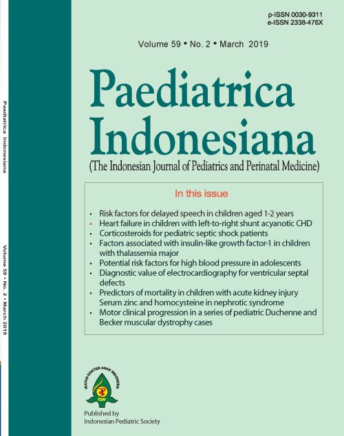
Diagnostic value of electrocardiography for ventricular septal defect
Abstract
Background Congenital heart disease (CHD) in children requires attention from medical practitioners, because CHDs that are diagnosed early and treated promptly have good prognoses. Ventricular septal defect (VSD) is the most common type of congenital heart disease.
Objective To compare the accuracy of electrocardiography (ECG) to echocardiography in diagnosing VSD.
Methods This diagnostic study was conducted from November 2013 until July 2015. It involved patients with acyanotic CHDs who were suspected to have VSD at Dr. Wahidin Sudirohusodo Hospital, Makassar, South Sulawesi.
Results Of 114 children screened, 97 were included and analyzed. The frequency of positive VSD was 69.1% based on ECG, and 99% based on echocardiography. There was a significant difference between ECG and echocardiography (P=0.000). However, when small VSDs were excluded, there was no significant difference between the two diagnostic tools [(P=1.000), Kappa value was 0.66, sensitivity was 98.5%, specificity was 100%, positive predictive value (PPV) was 100%, and negative predictive value (NPV) was 50%].
Conclusion There were significant differences between the ECG and echocardiography, for diagnosing VSD. However, if small VSDs were not included in the analysis, there was no difference between the two examinations, suggesting that ECG might be useful for diagnosing VSD in limited facilities hospitals.
References
2. Bernstein D. Acyanotic congenital heart disease: Kliegman, Berhman, Jenson, Stanton, editors. Nelson textbook of pediatrics. 19th ed. Philadelphia: Saunders Elsevier. 2011. p.1571
3. Meberg A, Otterstad JE, Froland G, Lindberg H, Sorland SJ. Outcome of congenital heart defects- a population based study. Acta Paediatr. 2000;89:1344-51.
4. Cahyono A, Rachman MA. The cause of mortality among congenital heart disease patients in Pediatric Ward, Soetomo General Hospital (2004-2006). Indones J Kardiologi. 2007;28:279-84.
5. Marelli AJ, Mackie AS, Ionescu-Ittu R, Rahme E, Pilote L. Congenital heart disease in the general population: changing prevalence and age distribution. Circulation. 2007;115:163-7.
6. Shah GS, Singh MK, Pandey TR, Kalakheti BK, Bhandari GP. Incidence of congenital heart disease in tertiary care hospital. Kathmandu Univ Med J. 2008;6:33-6.
7. Hariyanto D. Profil penyakit jantung bawaan di instalasi rawat inap anak RSUP Dr. M. Djamil, Padang Januari 2008 – Februari 2011. Sari Pediatri. 2012;14:152-7.
8. Rahayuningsih SE. Hubungan antara defek septum ventrikel dan status gizi. Sari Pediatri. 2011;13:137-41.
9. Shrivastava S. Malnutrition in congenital heart disease. Indian Pediatr. 2008;45:535-6.
10. Fyler DC, Geggel RL. History, growth, nutrition, physical examination and routine laboratory test. In: Keane FJ, Lock JE, Fyler DC, editors. Nada’s pediatric cardiology. 2nd ed. Philadelphia: Elsevier Saunders; 2007. p. 129-44.
11. Kimball TR, Daniels SR, Meyer RA, Hannon DW, Khoury P, Schwartz DC. Relation of symptoms to contractility and defect size in infants with ventricular septal defect. Am J Cardiol. 1991;67;1097-1102.
Copyright (c) 2019 Besse Sarmila, Burhanuddin Iskandar, Dasril Daud

This work is licensed under a Creative Commons Attribution-NonCommercial-ShareAlike 4.0 International License.
Authors who publish with this journal agree to the following terms:
Authors retain copyright and grant the journal right of first publication with the work simultaneously licensed under a Creative Commons Attribution License that allows others to share the work with an acknowledgement of the work's authorship and initial publication in this journal.
Authors are able to enter into separate, additional contractual arrangements for the non-exclusive distribution of the journal's published version of the work (e.g., post it to an institutional repository or publish it in a book), with an acknowledgement of its initial publication in this journal.
Accepted 2019-03-29
Published 2019-03-29


