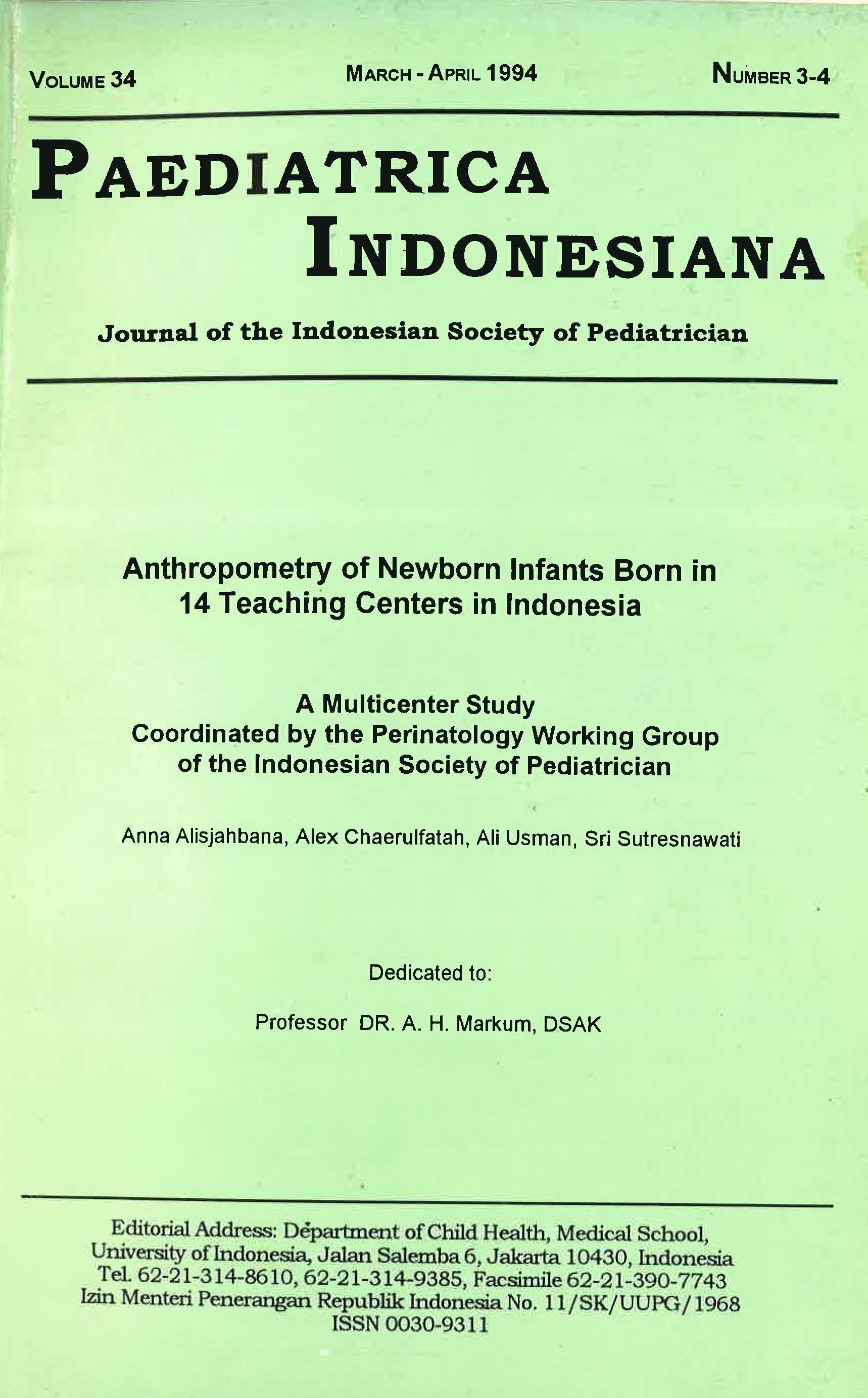
Anthropometry of Newborn Infants Born in 14 Teaching Centers in Indonesia
Abstract
Percentile curves representing intrauterine growth of Indonesian infants ranging from 34 to 43 weeks of gestation in 14 teaching centers were constructed from birth weight, birth length, and head, mid-upper arm, and chest circumferences. The gestational age was determined based on the last menstrual period. Mothers with probable chronic diseases or pregnancy complications were excluded. Included for analysis were 5844 singleton newborns. The mean birth weight of Indonesian babies was higher for gestational age of 34-38 weeks, but lower at 40-42 weeks of gestation compared with that of the Denver study. The results showed that the mean birth weight of Denver's newborns was significantly different than that of the Indonesian infants, therefore the Denver intrauterine growth curve cannot be used as reference curve for Indonesian newborns. Baby boys in general bad a higher mean birth weight, birth length, head circumference, and chest circumference. No difference was found for arm circumference. For every gestational age and percentiles, later born infants were heavier than first born infants. Birth weight at 42 weeks was lower for first born infants, this was not shown in later-born infants which showed higher weight for each percentiles. Parity affected birth weight more than birth length. Birth length became more stable at 39 weeks. Chest circumference of < 29 em had the highest sensitivit,y and positive predictive value for low birth weight, followed by arm circumference of < 9 cm. The use of intrauterine growth chart in studying the nutritional status of babies at birth was described.
References
2. Lechtig A., Sterky G, Tafari N. In birthweight Distnbution an Indicator of Social Development Sarec Report no. R:2; 1978, 87-90
3. Diamond r, Guidotti R. The use of a simple anthropometric measurements for predicting birth wejght Child Survival, Rearch Note Number 26 CS, 22 1969, June 2-10
4. Finstrom O. Studies on maturit;y in newborn infapts, birthweight, crown-heel length, head circumference and skull diameter in relation to gestational age. Acta P.ediatr Scand 1971; 60:685-94.
S. Miller HC, Merritt TA. Fetal growth in humans. Chicago: Yearbook Medical Publisher, 1979;31-82.
6. Birth weight surrogates. The relationship between birth w~t, ann and chest circumference. World Health Organization MCH/87.8
7. Rooth G, Meirik 0, Karlberg P. Estimation of the "normal growth of swedish infants at term", Preliminary Report Acta Pediatr Scand 1985; 319 (Supp1):76-9.
8. Petros-Barvazian, Behar M. problem identification. low birth weight-a major global problem. In: Birthweight distnbution an indicator of social development Sarec Report no. R:2, 1978., 9-15.
9. Puffer RR, Serrano CV. Patterns of birth weight, Pan American Health Organization Scientific Publication no. 504'. 1987;1-9.
10. Gruenwald P, Minh HN. Evaluation of body and organ weights in perinatal pathology. J Obstet Gynec 1961; 82: 312.
11.Mc Keown T, Record RG. The influence of placental size on fetal growth in man with special reference to multiple pregnancy. J Endocrinol1953; 9:418.
12. Falkner F. Key issues in perinatal growth. Acta Pediatr Scand 1985, 319 (Suppl) : 21-5.
13. Dwm PM. A Perinatal Growth Chart for International comparison. Acta Pediatr Scand 1985; 319 (Supp~180-7.
14. Ray Yip. Altitude and birth weight J Pediatr 1987,869-76.
15. Nishida H, Sakamoto S, Sakanoue M. New fetal growth cwves for Japanese. Acta Pediatr Scand 1985; 319 (Supp1):62-7.
16. Keen DV, Pearse RG. Intrauterine growth curves: problems and limitation. Acta Pediatr Scand 1985 (Suppl) 319:52-4.
Copyright (c) 2018 Anna Alisjahbana, Alex Chaerulfatah, Ali Usman, Sri Sutresnawati

This work is licensed under a Creative Commons Attribution-NonCommercial-ShareAlike 4.0 International License.
Authors who publish with this journal agree to the following terms:
Authors retain copyright and grant the journal right of first publication with the work simultaneously licensed under a Creative Commons Attribution License that allows others to share the work with an acknowledgement of the work's authorship and initial publication in this journal.
Authors are able to enter into separate, additional contractual arrangements for the non-exclusive distribution of the journal's published version of the work (e.g., post it to an institutional repository or publish it in a book), with an acknowledgement of its initial publication in this journal.
Published 2018-11-01

