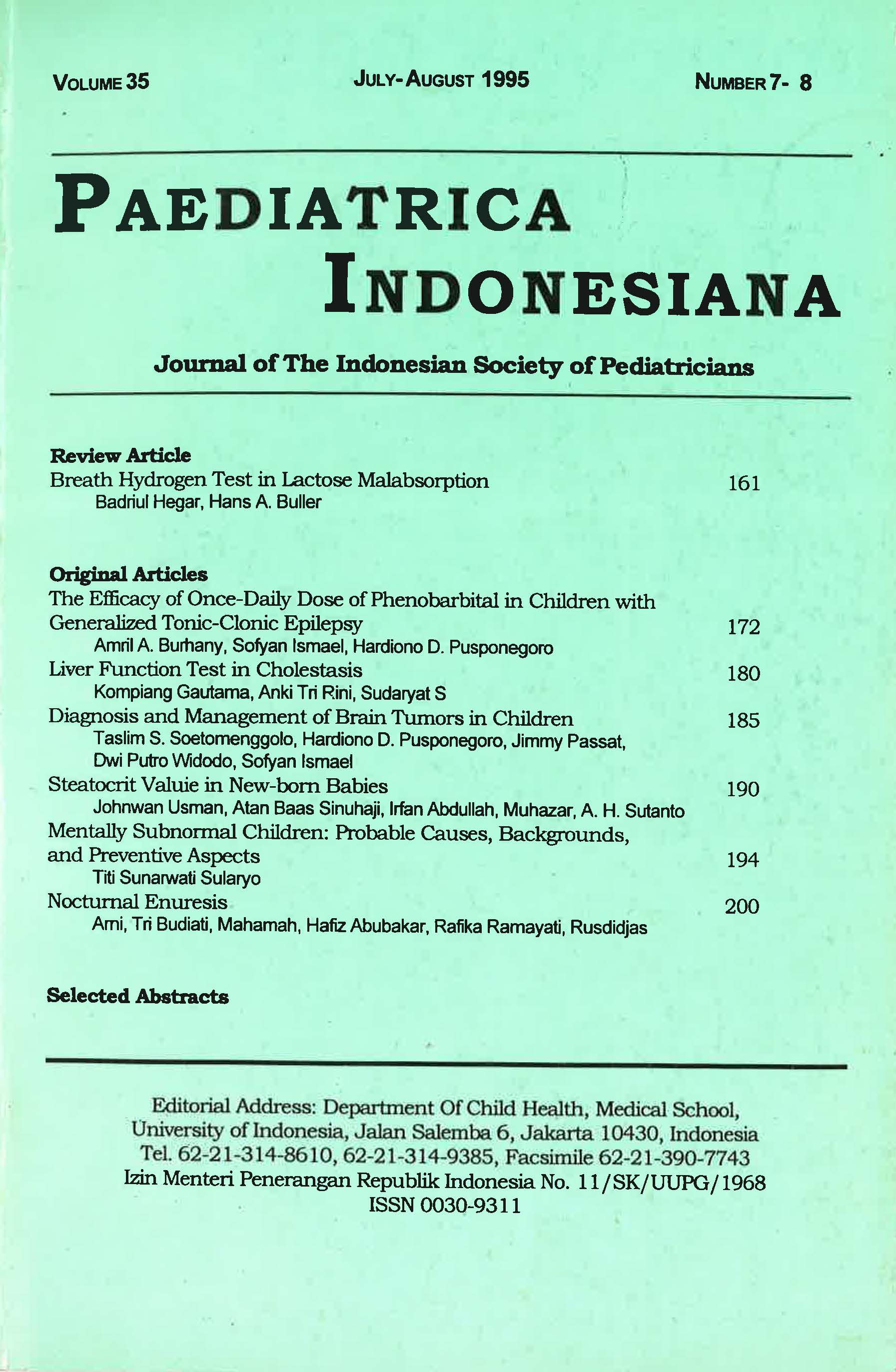
Liver Function Test in Cholestasis
Abstract
Cholestasis is impaired bile flow that cause prolonged evacuation of conjugated bilirubin and other substances which are dependent of bile flow for its excretion. The liver function test is useful to determine the severity of disease, to follow up its progress, and to predict the prognosis. This study was performed restropectively from the medical record of cholestatic patients who were admitted to the Department of Child Health, Central Hospital of Denpasar, from January 1992 to December 1993. Among 34 patients with cholestasis, 27 (19 intrahepatic and 8 extrahepatic cholestasis) were included in this study. Although the means of transaminase enzymes (SGOT, SGPT) in intrahepatic cholestasis were higher significantly than those in extrahepatic cholestasis, the increase of these enzymes five times or more than normal was not different significantly. The means of GGT and alkaline phospatase (AP) in extrahepatic groups were higher significantly than those in intrahepatic groups, and the increase of GGT more five times than normal was dilferent significantly as well. The means of total and conjugated bilirubin levels were higher in .extrahepatic group, but were not dilferent significantly.
References
2. Balistreri WF. Neonatal cholestasis. J Pediatr1985; 106:171-84.
3. Moyer MS, Balistreri WF. Prolonged neonatal obstructive Jaundice. In: Walker et al, eds. Pediatric gastrointestinal disease, pathofisiology-management. Vol 2. Philadelphia: BC Decker Inc, 1991; 835-7.
4. Boerhan H. Aspek klinis kolestasis pada anak. In: Erwin S, ed. Continuing education ilmu kesehatan anak, Hepatologi Anak FK Unair/RSUD Dr. Soetomo Surabaya. Surabaya, 1986; 53-63.
5. Purnamawati SP. Upaya diagnostik kolestasis pada bayi. In: Adnan SW, Zuraida Z, Pumamawati SP, eds. Hepatologi anak masa kini, Jakarta: UI Pres, 1992; 11-25.
6. Mowat AP. Laboratory assessment of hepatobiliary disease. In: Liver disorder in childhood; 2nd ed. London: Butterworths, 1987; 366-73.
7. Sherlock S. Assessment of liver function. In: Disease of the liver and biliary system. Boston: Blackwell Scientific Publ, 1989: 19-35.
8. Wijaya A. Diagnosis laboratorik penyakit hati. In: Program Pustaka Prodia, Seri Hepatitis 03, Bandung; 1990.
9. Halimun EM. Current management on neonatal obstructive jaundice. Pediatr lndones 1984; 24:101-3.
10. Halimun EM, Maryana. Penanganan kholestasis pada bayi In: Erwin S, ed. Continuing Education, llmu Kesehatan Anak, Bedah Anak, FK Unair/RSUD Dr. Soetomo Surabaya, 1984; 77-83.
11. Ferry GO, Selby ML, Udall J, et al. Guide of early diagnosis of biliary obstruction in infancy, review of 143 cases. Clin Pediatr 1985; 24:305-11.
12. Koop CE. Progresive extrahepatic biliary obstruction of the newborn. J Pediatr Surg 1975; 10:169-70.
13. Wiharta AS. Hati dan saluran empedu. In Markum AH et al, eds. Buku ajar ilmu kesehatan anak. Jakarta: Ul Press, 1991; 513-22.
14. Balistreri WF, Suchy FJ, Ferre MK, Heubi JE. Pathologic versus physiologic cholestasis: Elevated serum concentration of secondary bile acid. J Pediatr 1991; 98:399-402.
Copyright (c) 2018 Kompiang Gautama, Anki Tri Rini, Sudaryati S.

This work is licensed under a Creative Commons Attribution-NonCommercial-ShareAlike 4.0 International License.
Authors who publish with this journal agree to the following terms:
Authors retain copyright and grant the journal right of first publication with the work simultaneously licensed under a Creative Commons Attribution License that allows others to share the work with an acknowledgement of the work's authorship and initial publication in this journal.
Authors are able to enter into separate, additional contractual arrangements for the non-exclusive distribution of the journal's published version of the work (e.g., post it to an institutional repository or publish it in a book), with an acknowledgement of its initial publication in this journal.
Published 2018-10-08

