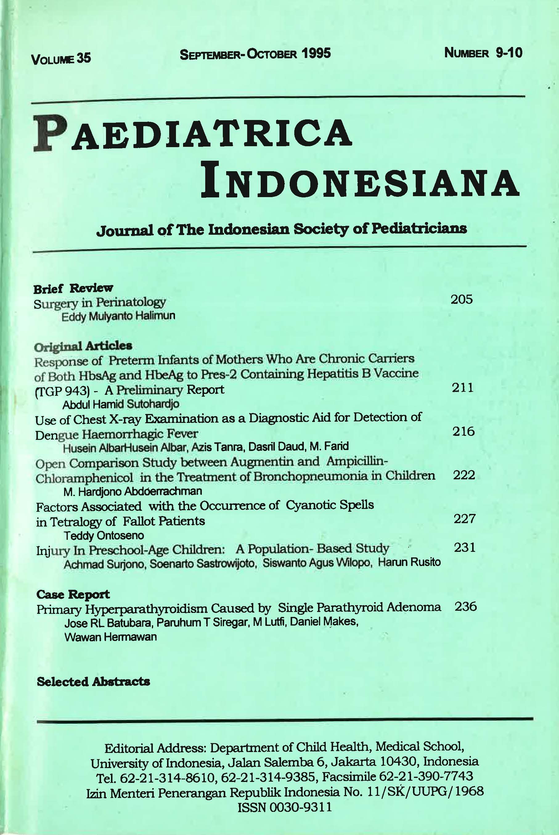
Use of Chest X-Ray Examination as a Diagnostic Aid for Detection of Dengue Haemorrhagic Fever
DOI:
https://doi.org/10.14238/pi35.9-10.1995.216-21Keywords:
chest xray; dengue haemorrhagic fever; RLD position; pleural effusionAbstract
The advantage of a chest X-ray in the RLD position in 15 children with DHF hospitalized at the paediatric ward of the Ternate General Hospital within the period of May-June-July 1990 and June-July 1991 were evaluated. Besides clinical and laboratory assessment to establish the diagnosis of DHF according to the WHO guidelines (1975), child and a haemaglutination-inhibition test was also done to confirm the diagnosis.
Chest X-rays in the RLD position found a pleural effusion in 11 out of 15 patients with DHF especially in those with dengue shock syndrome. A positive serological test was always associated with the presence of PE (100%), while this could be shown in only 2 patients with negative test results. It may be concluded that the WHO criteria for the clinical diagnosis of DHF may be confirmed not only by the serological test but also by the presence of PE on chest film in the RLD position and therefore this examination may play an important role in establishing a diagnosis of DHF in a regency hospital.
References
2. Suroso T. Status epidem.iologi dan strategi pemberantasan demam berdarah dengue. Naskah Lengkap Pendidikan Tambahan Berkala llmu Kesehatan Anak. Demam Berdarah (Dengue) Jakarta,
1982:74-78.
3. Sumarmo. Demam berdarah dengue pada anak. Tesis U1 1983. Penerbit Universitas Indonesia (VI-Press).
4. Tamaela LA dan Karjomanggolo HWT. Peranan pemeriksaan radiologik toraks pada dengue baemorrbagic fever. Nama lengkap pendictikan tambahan berkala ilmu kesehatan anak. Demam berdarah (dengue), 23 Januari 1982.
5. Tarau JL, Azis Tanra, Dasril Daud. Efusi pleura pada demam berdarah dengue. Naskah lengkap Pendidikan spesialisasi ilmu kesehatan anak. FKUH, 1987.
6. Sumarmo. Demam berdarah dengue : Aspek klinis dan penatalaksanaan. CDK. 1990;60:11-5.
7. Tanra A, Makaliwy Ch. Pemeriksaan radiologic pada demam berdarah dengue. Simposium demam berdarah dengue. Ujung pandang, 1989.pP. 11-5.
8. Sunoto. Demam berdarah dengue: Sepuluh tahun penelitian pada anak di Jakarta. Jakarta, 1985. p. 37-9.
9. Meschan I. Synopsis of analysis of roentgen signs in general radiology. Philadelphia, London, Toronto: WB Saunders Co; 1969. p. 13-53.
10. Gyn C, Blake N. Pediatric diagnostic imaging. London: William Heinemann Medical Books; 1986. p. 193-7.
Downloads
Published
How to Cite
Issue
Section
License
Authors who publish with this journal agree to the following terms:
Authors retain copyright and grant the journal right of first publication with the work simultaneously licensed under a Creative Commons Attribution License that allows others to share the work with an acknowledgement of the work's authorship and initial publication in this journal.
Authors are able to enter into separate, additional contractual arrangements for the non-exclusive distribution of the journal's published version of the work (e.g., post it to an institutional repository or publish it in a book), with an acknowledgement of its initial publication in this journal.

