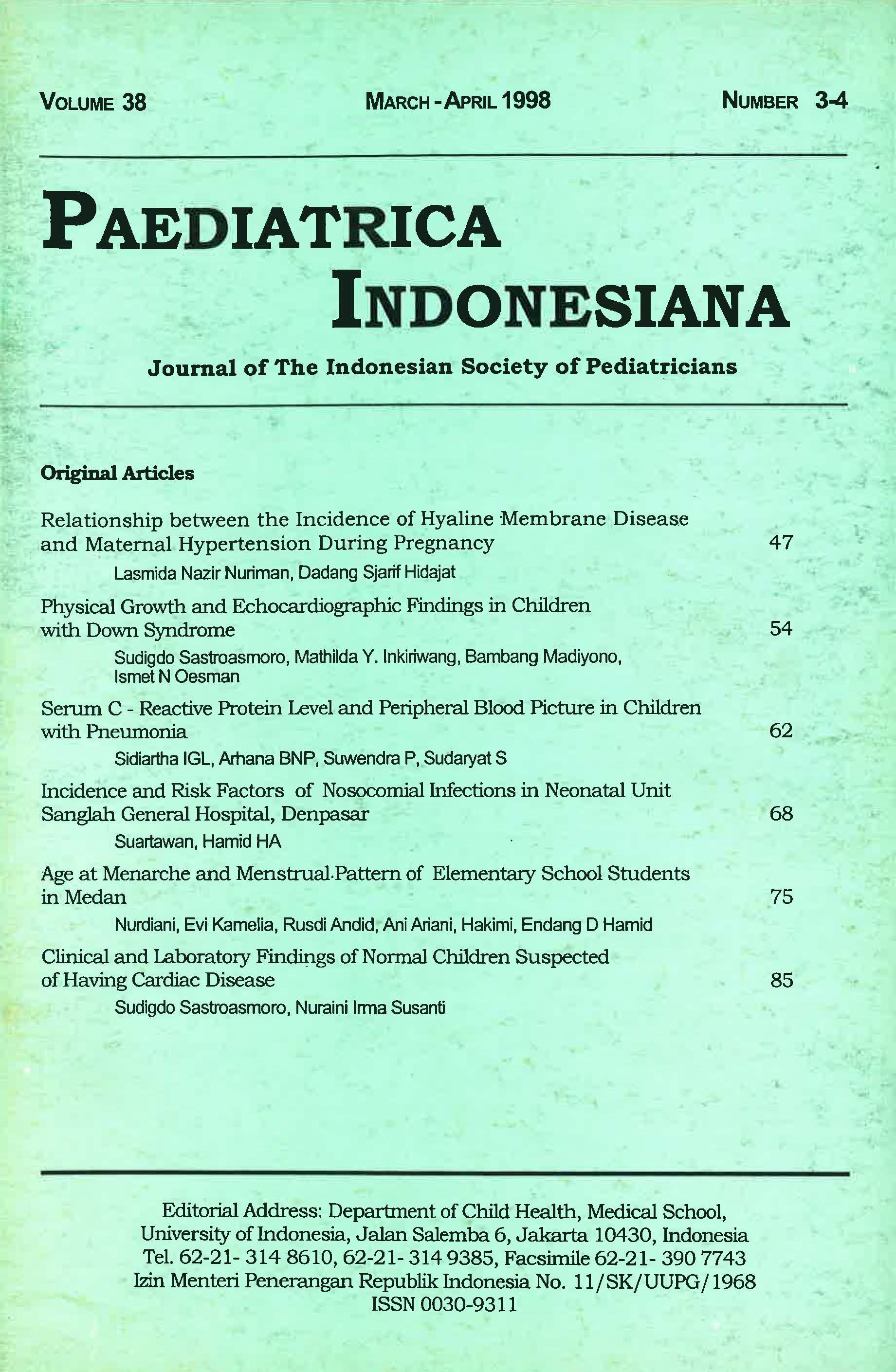
Physical Growth and Echocardiographic Findings in Children with Down Syndrome
Abstract
We compared the physical growth, nutritional status, and echocardiographic findings in children aged 3-7 years with Down syndrome who had no congenital heart disease. Thirty such patients who consecutively referred to the Division of Cardiology, Department of Child Health, Medical School, University of Indonesia/Cipto Mangunkusumo Hospital, Jakarta, were compared with sex and age matched controls consisted of normal children attending the Department. It appears that growth and nutritional status of children with Down syndrome tended to be retarded when compared to those of the controls. However, no significant difference were found on the M-mode echocardiographic values of the left ventricle, except that the left ventricular posterior wall thickness in study subjects was more that that of the controls. We concluded that although the pulmonary architecture of patients with Down syndrome is thought to be less developed than that of normal children, it does not affect the left ventricular measurements and function as measured by M-mode echocardiography.
References
Kadri N, Siregar SP, Surachman HS, Monintja HE. Umur ibu sebagai faktor risiko kelahiran bayi mongoloid d1 Rumah Sakit Cipto Mangunkusumo, Jakarta, 1975-1979. Maj Obstet Ginekol Indones 1982; 8:147-54.
Mikkelsen M. Down's syndrome. Current stage of cytogenetic research. Hum Genet 1971;12:1-12.
Lindsten J , Marsk L, Berlind KS, et al. Incidence of Down's syndrome in Sweden during the years 1968-1977. ln: Burgio R. Trisomy 21. Hum Genet (Suppl2) 1981; 195-210.
Spicer RL. Cardiovascular disease in Down's syndrome. Pediatr Clin North Am 1984;31:1331-43.
Rowe RD, Uchida IA. Cardiac malformation in mongolism. Am J Med 1961; 31:726-35.
Tandon R, Edwards JE. Cardiac malformation associated with Down's syndrome. Circulation 1973; 47:1349-55.
Sugiyama H. Property of myocardium in Down syndrome without congenital heart disease. J Japan Pediatr 1991;1340-5.
Cooney TP, Thurlbeck WM. Pulmonary hypoplasia in Down's syndrome. N Engl J Med 1982; 307:1170-3.
Hook EB. Down syndrome. Frequency in human population and factors pertinent to variation in rates. In de la Cruz G. Trisomy 21 (Down syndrome). Baltimore: University Park Press, 1981 ;8-6 7.
Ramelan W. Personal communication.
Caffey's pediatric X-ray diagnosis: an integrated imaging approach. 9th ed. St Louis: Mosby; 1993.
Roelandt JRTC, Sutherland GR, Iliceto S, linker DT. Cardiac ultrasound. Edinburgh:Churchill Livingstone; 1993.
Berg JM, Crome L, France NE. Congenital cardiac malformation in mongolism. Brit Heart J 1960; 22:331-46.
Anneren G, Gustavson KH, Sara VR. Growth retardation in Down's syndrome with special reference to somatomedin. The 7th Congress of IAASSMD. New Delhi, India, 24-28 March 1985.
Authors who publish with this journal agree to the following terms:
Authors retain copyright and grant the journal right of first publication with the work simultaneously licensed under a Creative Commons Attribution License that allows others to share the work with an acknowledgement of the work's authorship and initial publication in this journal.
Authors are able to enter into separate, additional contractual arrangements for the non-exclusive distribution of the journal's published version of the work (e.g., post it to an institutional repository or publish it in a book), with an acknowledgement of its initial publication in this journal.
Accepted 2017-07-06
Published 2017-07-11

